
Microscope dentistry — when the smallest detail matters
The aim of modern dentistry is no longer to extract and replace a damaged or dead tooth as soon as possible — on the contrary: we want to save as much of the patient's own tooth structure as possible.
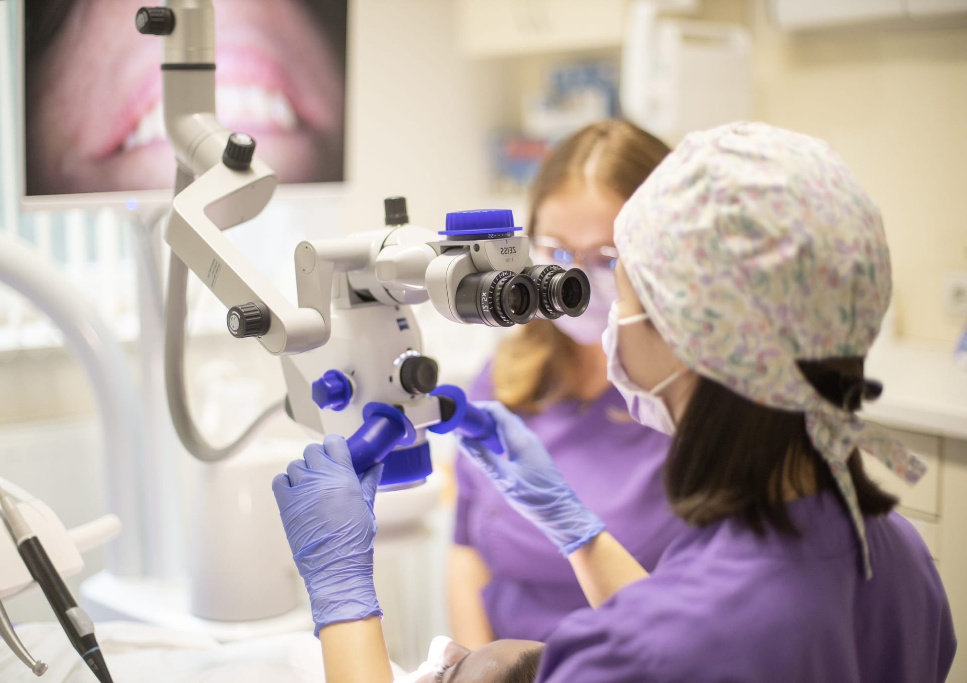
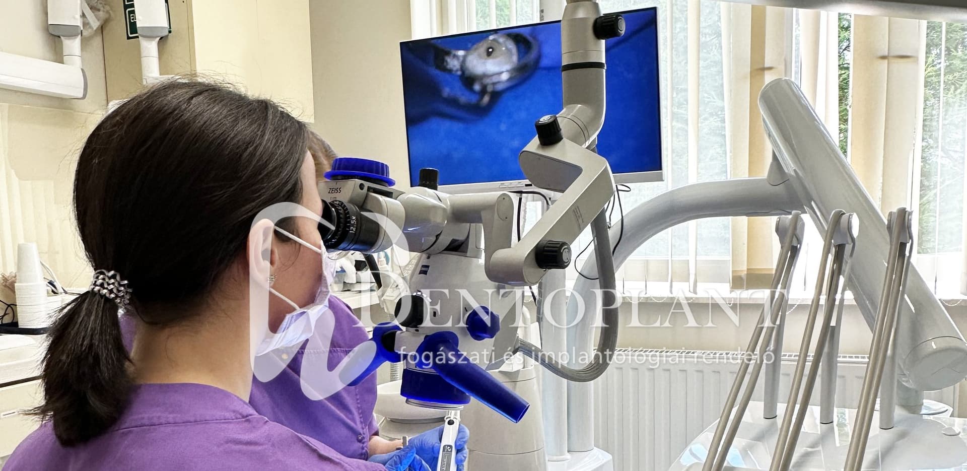
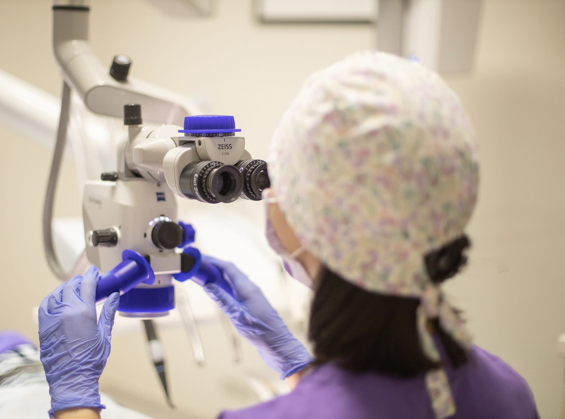
One of the most important tools for this is the operating microscope, which provides up to 25× magnification, so we can see details that would not be visible to the naked eye or even with loupes.
At the Dentoplant Dental Clinic we use one of the world's leading microscopes, the Zeiss Extaro 300.
Treatment takes place with the patient lying down, isolated with a rubber dam. The microscope image is also visible on a monitor.
Why does magnification matter in dentistry?
The advantage of magnification during dental treatment is beyond dispute, especially in root canal treatment, where success often depends on tenths of a millimetre. Magnification is indispensable because:
- we see the tiny details of the tooth much more accurately
- complications are easier to avoid
- the internal anatomy of the tooth is easier to detect, which speeds up treatment
- the procedure is safer and more predictable
While loupes typically give 2.5–6× magnification, the microscope approaches the area at 16–25×, so we can work with better visual control. Its intense, shadow-free light makes every detail clearly visible. An extra function is fluorescent lighting: carious areas appear in a different colour, so they can quickly be distinguished from healthy tooth structure.
What do we use the microscope for?
1. Root canal treatment
It is especially useful when:
- the root canal is very curved or narrow
- an extra canal or deeper branches need to be found
- a previous treatment has left a preparation error or obstruction
- retreatment is needed (replacing inadequate root fillings that often do not reach the apex and are not wall-tight, then proper preparation and filling)
The microscope then helps us see the canal system precisely and solve the problem with minimal removal of tooth structure. Our clinic also accepts patients referred specifically for root canal treatment by external colleagues.
2. Removing a broken root-canal instrument
A root-canal file can break inside the tooth, typically in narrow, curved canals. With the microscope we can:
- locate the fragment
- prepare around it safely
- remove it with special techniques — this would be impossible without a microscope
3. Root-end surgery (resection)
Magnification also allows us to work precisely in surgical procedures.
4. Diagnostics
- early caries
- crack lines
- misfit of fillings and restorations
- root-canal anomalies
- certain gum and oral-mucosa changes
5. Preparing precision restorations
When preparing fillings, inlays and veneers, the marginal seal can be formed much more finely under magnification.
6. Periodontal (gum) surgery
- in tooth-preserving pocket treatment, the cleanliness of the bone wall can be checked
- in cyst removal, completeness of removal can be assessed
- in gum-recession surgery
- when placing microsurgical sutures
What happens during a microscope treatment?
The treatment is comfortable, safe and completely painless. The patient is lying down, so the procedure takes place in a more relaxed, stable position, and we have a better view of the area. The tooth is isolated with a rubber dam (cofferdam), which protects the patient and keeps the field clean. The microscope image is also visible on a monitor, so the patient can see what is happening during treatment.
Removing a broken instrument — a real case
Our colleague Dr Eszter Sebők presents a common yet special situation: how a previously broken root-canal file that caused inflammation and pain can be removed.
1. Starting condition
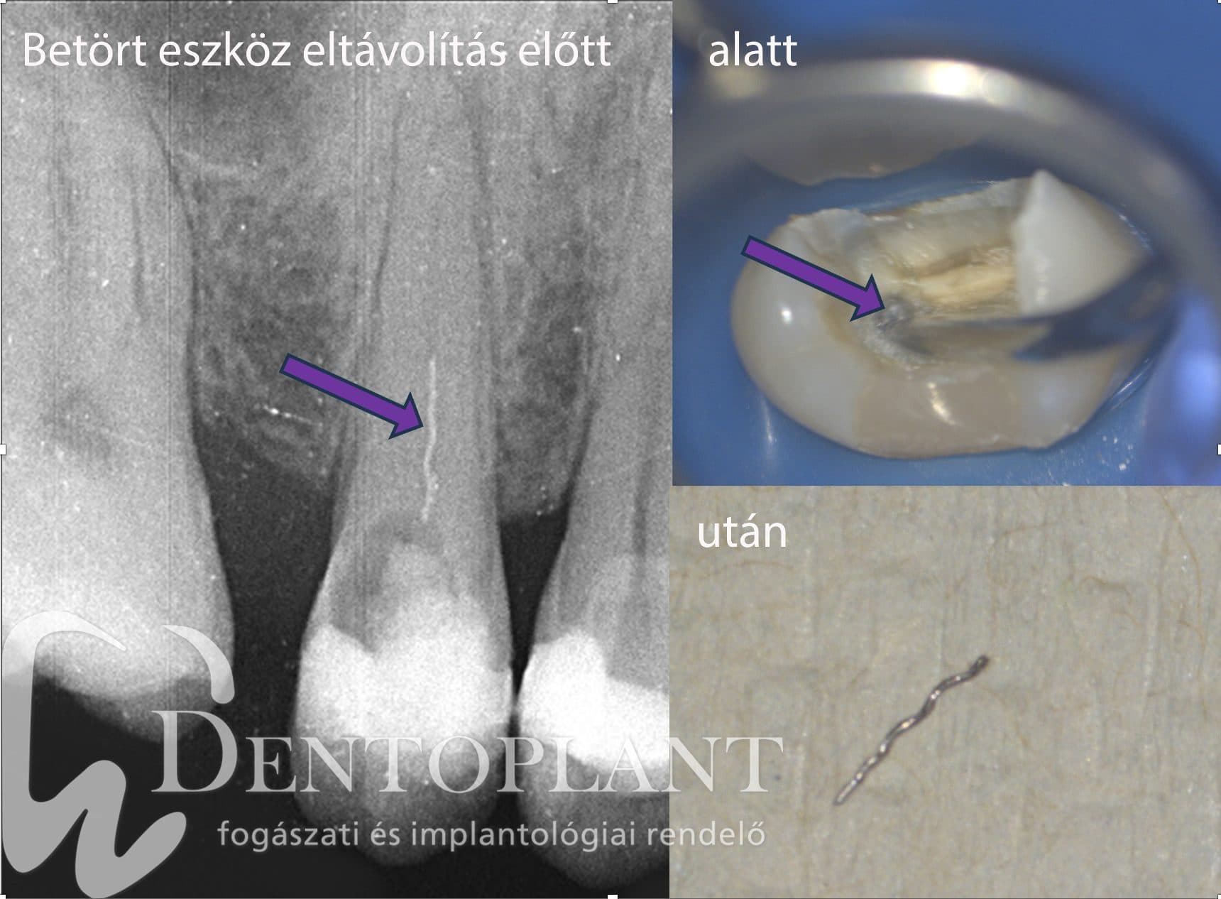
The patient arrived in severe pain. Their dentist had started root canal treatment but could not continue because the narrow canals were impassable, and referred them for specialist care. The X-ray clearly showed a broken file remaining in one canal of a premolar, which made disinfection impossible.
2. Microscopic exposure
After proper isolation we removed the temporary filling under the microscope — the instrument was already visible in one canal. We started to move the fragment with circular ultrasonic vibration, then rotated it out of the canal. Treatment did not end there: under microscopic magnification we also opened the difficult, narrow canals.
3. Result
The fragment came out successfully, confirmed by a control X-ray, and both canals became passable along their full length.
This case shows that the microscope is often the key to saving the tooth.
During treatment
In summary
Microscope dentistry is one of the most modern and precise technologies today. It:
- 1increases the success of treatments
- 2helps avoid unnecessary extractions
- 3is gentle and safe
- 4gives the patient a better experience